Case Report: Corneal Perforation after CXL
Corneal Collagen Cross-Linking: Are there Unknown Contraindications?
ROLES:
Qualitative Researcher, Paper Writer
Research Overview: Corneal Collagen Crosslinking
Corneal collagen crosslinking (CXL) is a newer treatment for keratoconus. It is believed that the pathophysiology of this disease may be in part related to variations in how collagen in the cornea is remodeled following mechanical stress. Over time, a normal ocular pressure on a weakened surface causes alterations in shape. In CXL, riboflavin is instilled into the corneal stroma and irradiated with UV light, causing the formation of free-radicals, which interact with nearby collagen molecules to form new molecular bonds. It has been shown that this stiffens the cornea and effectively slows progression of the disease. The complication rate of the procedure is very low when following guidelines.
For this reason, we were surprised to have seen a patient who was diagnosed with keratoconus and subsequently had epithelium-off crosslinking, only to spontaneously have a corneal perforation following minimal trauma.
Working with Yaron Rabinowitz, M.D., Ron Gaster, M.D., and Deborah Jacobs, M.D., I performed a retrospective chart review on this patient. Realizing that a genetic analysis would be helpful in preventing this complication in the future, I involved Anthony J. Aldave, M.D., Wenlin Zhang, M.D., Ph.D..
What happened with this patient?
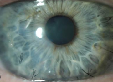
The patient, an Ashkenazi Jewish woman in her sixth decade, had rapidly progressing myopia in her teens which stabilized and was correctable with spectacles and RGP lenses. In her 50s, she developed topographic astigmatism which was not correctable with glasses and was diagnosed with keratoconus vs pellucid marginal degeneration. Based on the corneal tomographic findings, the patient was diagnosed with keratoconus and underwent epi-off CXL in the right eye. One month and four months after the CXL procedure, spontaneous micro-perforations developed in the right cornea. Slit-lamp examination after the second perforation demonstrated a diffusely thin cornea with a temporal paracentral perforation. Both perforations were managed conservatively with cyanoacrylate glue and bandage contact lens placement and aqueous suppression. One year after the second perforation, the vision in the right eye was correctable to 20/60 on refraction and 20/30 with a KeraSoft® IC lens.
The following year, the patient noted worsening of vision secondary to progressive myopia and astigmatism in the left eye. The topographic astigmatism had progressed to 4.7 D. Given the history of perforation following epi-off CXL for the right eye, epi-on CXL was performed for the left eye. However, eight days after the epi-on CXL, a spontaneous corneal perforation developed in the left eye, which was managed with sutures, amniotic membrane transplantation, and fibrin sealant. Seven weeks after CXL, following suture removal in left eye, a leak developed in the area of the initial perforation, which was managed conservatively. Nearly two years after the CXL was performed for the left eye, a spontaneous cornea perforation developed in the right eye that required suturing and amniotic membrane transplantation for closure. After recovery, the right eye was fit with a BostonSight® PROSE device, providing 20/40 CDVA. Review of systems and continued follow-up of the proband reveal no history or evidence of immunologic or rheumatologic conditions
Although the global corneal thinning extended to the limbus in the proband and her daughter, the corneal thickness profiles were within the 95% (± 2 SD) interval around the mean reported for keratoconic corneas at central, paracentral (4 mm) and peripheral (8 mm) locations (with the exception of the peripheral corneal thickness in the proband) with maximal thinning at the apex in both the proband and her daughter (Table 1).30, 31 Based on this, both the proband and her daughter were diagnosed with keratoconus as opposed to keratoglobus.
What was found on genetic analysis?
Using Sanger sequencing of ZNF469 in the proband, Dr. Zhang identified three rare non-synonymous single nucleotide variants (SNVs), all heterozygous (c.2035G>A, c.10244G>C, and c.11119A>G), and all with a MAF ≤ 0.01 (Table 2). Screening of ZNF469 in the proband’s affected daughter and the unaffected biological father of the daughter identified the same three variants in the heterozygous state in the daughter but not in the daughter’s father.
In silico analyses of the resulting amino acid substitutions, and while the most impactful of the three was predicted to be “possibly damaging,” the combined effect of the three missense variants cannot be predicted.
Why is this important?
Corneal cross-linking may be associated with the development of corneal perforation in particular at-risk individuals with keratoconus. Identifying clinical and genetic risk factors, including screening of ZNF469 and PRDM5, may be useful in the prevention of significant complications following CXL.
This case report was accepted for publication in Cornea.